The short answer
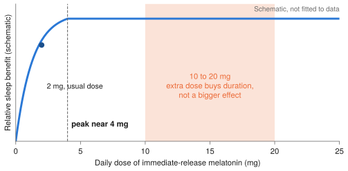
- Sleep: the benefit plateaus near 4 mg. A dose-response meta-analysis of 26 randomized trials found sleep-onset latency and total sleep time improve up to about 4 mg a day and no further.1 At 10 to 20 mg the extra dose mostly buys duration, not a larger effect.
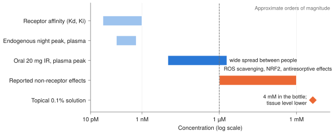
- The rest of the case lives at micromolar concentrations. Radical scavenging, NRF2 activation and much of the anticancer and neuroprotective work are reported at about 1 µM and above, several orders of magnitude over the nocturnal plasma level. One 2026 review argues that conventional oral dosing cannot sustain that exposure, and another group notes the receptors are desensitized in that range.6,8
- Cancer is the one signal at 20 mg, and it is weak. Older meta-analyses are strongly positive; the 2025 Cochrane review rates the evidence very low certainty, with fatigue the one outcome that probably improves.9,10,11
- Skin and hair: topical beats oral. The human data are almost all topical, and the mouse hair mechanism is receptor-dependent, the pathway a modest oral dose already occupies.15,16,17
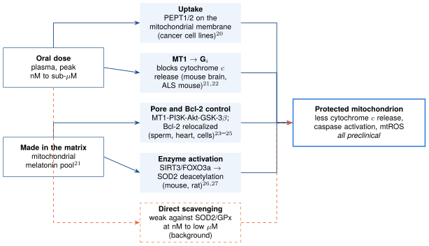
- Mitochondria are the strongest non-sleep case, but the protection looks mostly indirect: receptor signaling, Bcl-2 and permeability-pore control, and SIRT3-SOD2 activation, not stoichiometric radical scavenging. Everything is preclinical.20–27
| Outcome | Human evidence | What the mechanism rests on | Confidence |
|---|---|---|---|
| Sleep onset and duration | 26-RCT dose-response meta-analysis, plateau near 4 mg1 | MT1/MT2 signaling, well characterized | High at 0.5 to 4 mg; nothing shows added benefit at 10 to 20 mg |
| Cancer, adjuvant | Positive older meta-analyses at 20 mg; Cochrane 2025: very low certainty9,10,11 | Oncostatic, immune and antioxidant effects, mostly in vitro | Low |
| Neuroprotection | No disease-modifying effect shown (MS review); small sleep-driven gains4 | Mitochondrial and receptor signaling in rodents21,22 | Low |
| Longevity | None in humans | Antioxidant and circadian arguments | Very low |
| Skin, oral | No good oral trials found | Skin makes melatonin and expresses MT1/MT218 | Very low |
| Hair, topical | Pilot RCT; 8 of 11 studies positive15,16 | Wnt/β-catenin, receptor-dependent in mice17 | Low |
| Greying | None found | Melanocyte-stem-cell oxidative stress (background) | Very low |
Section oneWhat a bigger dose actually changes
The first question about 20 mg is what the extra 16 mg does. For a receptor-mediated hormone with nanomolar affinity, the answer is that it moves you off the steep part of the dose-response curve and onto the flat part, where more drug mostly extends how long the receptors stay occupied.
The dose-response meta-analysis is the cleanest evidence. Across 26 double-blind trials in healthy volunteers and people with insomnia, the effect on sleep-onset latency and total sleep time rose gradually and peaked at 4 mg a day. Timing mattered too: giving the dose about three hours before the desired bedtime predicted a bigger effect than the common 2 mg at 30 minutes.1 A separate pediatric neuropsychiatric meta-analysis reached a similar plateau, with maximal efficacy between 2 and 4 mg.2
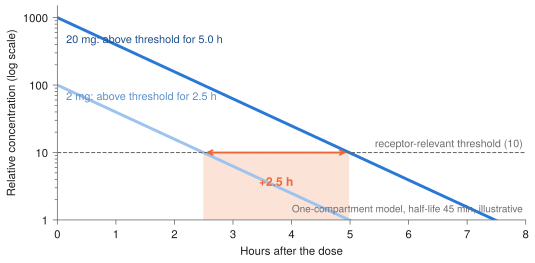
Pharmacokinetics explain why. The half-life of immediate-release melatonin is about 45 minutes, so a tenfold dose adds only about 3.3 half-lives (the base-2 logarithm of 10), roughly 2.5 hours more time above any fixed threshold. That is an extension of the same effect, not a stronger one.
Background knowledge, not retrieved
Oral bioavailability averages about 15%, with a large spread driven by CYP1A2. Inhibitors such as fluvoxamine and ciprofloxacin can raise exposure several-fold, and the enzyme induction from tobacco smoke disappears within weeks of quitting, so levels at a fixed dose can climb. Nicotine alone does not induce CYP1A2. At 10 to 20 mg this variance matters more than the dose itself.
Section twoSleep and the clock
The one signal in favor of higher doses comes from a small meta-analysis in Parkinson's disease: five randomized trials, 206 patients. Doses above 4 mg improved the Pittsburgh Sleep Quality Index by 2.90 points (95% CI 1.78 to 4.02) against 1.26 (not significant) for 4 mg or less, and immediate-release beat prolonged-release.3 That is a between-trial subgroup comparison in a single disease, so it is hypothesis-generating. Short treatment (4 weeks) worked; 8 to 12 weeks did not reach significance.
The less-groggy morning that some people report on IR 20 mg compared with 6 mg SR fits the kinetics: SR is built to hold levels into the morning, IR clears within hours. It is also the reason the dose-response data matter. If the benefit is saturated, the main thing a high IR dose changes is how long the signal persists.
Background knowledge, not retrieved
Melatonin shifts the circadian clock according to a phase-response curve: given in the evening it advances the clock, given in the early morning it delays it. A high dose that lingers into the morning can overlap the delay zone and blunt the advance, while 0.3 to 0.5 mg already advances the clock about as well. Commonly reported side effects at high doses are vivid dreams, morning grogginess, headache and a transient fall in core temperature. If maintenance is the goal, prolonged-release or a split dose extends exposure more cleanly than going to 20 mg IR.
Section threeReceptor biology and the exposure problem
MT1 and MT2 are high-affinity G protein-coupled receptors. Agonist exposure drives β-arrestin recruitment, after which the receptor is internalized and G-protein signaling stops; this is described directly for MT1.5 In rats, a hemorrhage-and-resuscitation model with 1 mg/kg intravenous melatonin or ramelteon showed a 50% fall in hepatic MT1 protein, possibly by desensitization, although hemorrhage itself also contributed.7 I found no human data showing tolerance to the hypnotic effect, so desensitization is a plausible concern, not a demonstrated one.
The sharper question is what happens at the other end. Many of melatonin's celebrated actions, direct antioxidant effects, NRF2 activation and antiresorptive effects, are reported at about 1 µM or more. A 2026 review puts these four to six orders of magnitude above nocturnal plasma levels, attributes them increasingly to receptor-independent chemistry shared with structurally unrelated antioxidants, and concludes that sustained exposure at or above 1 µM is not attainable by conventional oral dosing.8 Another group found no effect of melatonin in standard immuno-inflammation cell models and noted that the reported effects appear at 1 µM and beyond, where the receptors are desensitized by internalization.6 Note that Lin's review concerns bone and Boutin's concerns inflammation; I am applying the exposure argument by analogy to the other outcomes.
Section fourCancer
The positive trials behind the 20 mg figure are older and small. An eight-trial meta-analysis (761 patients), in which every trial used 20 mg orally alongside chemotherapy or radiotherapy, reported a one-year survival risk ratio of 1.90, higher remission rates and less thrombocytopenia, neurotoxicity and fatigue.9 A 21-trial analysis found a one-year mortality risk ratio of 0.63.10 An early 80-patient randomized pilot at 20 mg a day found less thrombocytopenia and less malaise and asthenia, with no effect on alopecia or vomiting.14
The 2025 Cochrane review is the counterweight. It covers 30 randomized trials and 5,093 participants, with 28 at high risk of bias. Survival was not established; fatigue probably falls (RR 0.46, moderate certainty) and nausea may fall (low certainty), and it concludes that no firm decision for or against adjunctive use can be made.11 A 19-trial meta-analysis found no effect on quality of life, sleep, fatigue or pain.12 For prevention, a small meta-analysis linked low melatonin to breast cancer, but that is observational and does not show that supplementation prevents anything.13
How I read it
Twenty milligrams is the dose behind the positive trials, but the trials are small, heterogeneous and largely at high risk of bias. That makes the survival signal a hypothesis, not an effect. It is evidence for patients already on treatment, not for prevention in healthy people, and it is not a substitute for any therapy.
Section fiveNeuroprotection and longevity
The preclinical case is strong: melatonin supports mitochondrial function and reduces apoptosis and neuroinflammation in many rodent models (the mitochondrial section below covers what that rests on). The translation problem is dose. Rodent studies commonly use 10 to 100 mg/kg, while 20 mg in an adult is about 0.3 mg/kg, and the micromolar brain exposure behind many in vitro effects is out of reach orally.
The human evidence is mostly indirect. A multiple-sclerosis systematic review of 10 studies (435 participants, doses 0.5 to 300 mg a day) found no reduction in relapse, disability progression or new MRI activity, and rated certainty low or very low for every outcome. Small studies hinted at gains in sleep, fatigue and mood. Doses of 0.5 to 25 mg a day were generally tolerated; at 300 mg a day three of four participants developed raised transaminases, which recurred on rechallenge in one.4 Beyond that, an early 3 mg migraine report exists,29 and from background knowledge, delirium prevention trials are small and mixed and a 300 mg a day rectal ALS trial showed safety but no efficacy.
For longevity, rodent lifespan results are inconsistent and I know of no human lifespan or healthspan data. Any plausible human benefit runs through sleep quality and circadian alignment. A 2024 review claims anti-aging effects across brain and skin, but it is a narrative review of mechanisms and preclinical work.19
Section sixSkin, hair, greying and visible aging
Skin. Skin synthesizes melatonin and expresses its receptors, and melatonin has antioxidant, DNA-repair and UV-protective properties there.18 I found no good randomized trials of oral melatonin for wrinkles, elasticity or collagen, though my searches here were noisy. From background knowledge, topical application before UV exposure reduced erythema in small human studies. Benefit from systemic dosing is largely inferred, and better sleep may be the more realistic route to better-looking skin.
Hair loss. In a double-blind pilot, 0.1% topical melatonin applied daily for six months to 40 women raised anagen rate in occipital hair in androgenetic alopecia (n = 12, P = 0.012) and in frontal hair in diffuse alopecia (n = 28, P = 0.046), with plasma melatonin staying below the physiological night peak.15 A 2023 review found 11 human studies, eight with positive results for topical use in androgenetic alopecia, at 0.0033% to 0.1% once daily for 90 to 180 days, versus 1.5 mg orally twice daily.16 In mice, melatonin promoted regeneration through Wnt/β-catenin signaling in dermal papillae and follicle stem cells, and the receptor antagonist luzindole suppressed it, pointing to a receptor-dependent mechanism.17 Oral 20 mg did not prevent chemotherapy-induced alopecia.14
Greying. My search returned nothing usable. Background biology says pigment loss reflects melanocyte-stem-cell depletion and oxidative stress, which makes melatonin a plausible in vitro protector, but I know of no human trial.
Practical read
If hair or skin is the goal, topical melatonin has more human data than raising the oral dose. Because the hair mechanism looks receptor-mediated, I would not expect much extra from going above a few milligrams by mouth.
Section sevenIs mitochondrial protection direct radical scavenging?
Partly, but probably not mainly. Mitochondria are the best non-sleep case for an oral dose, and the literature I retrieved points to a mix of local accumulation, receptor signaling inside the organelle, and enzyme activation, with direct scavenging contributing an unknown and probably minor share.
How melatonin might get in, and what it does when it does
Uptake. In human cancer cell lines (PC3 and U118), PEPT1 and PEPT2 localized to the mitochondrial membrane and carried melatonin into mitochondria, where its accumulation induced apoptosis.20 That is the only paper my search returned for this question, from one group, in cancer cell lines, and I read only the abstract. It shows a transport route exists in those cells; it does not show human tissue concentrations.
The receptor inside the organelle. Suofu and colleagues found MT1, its G protein and β-arrestins on and within neuronal mitochondria, and reported that melatonin is synthesized in the mitochondrial matrix and released to activate that pool of MT1, which inhibits stress-induced cytochrome c release and caspase activation. Overexpressing mitochondrial MT1 reduced ischemic brain injury in mice.21 They call this "automitocrine" signaling. In a mouse model of ALS, melatonin blocked mitochondrial cytochrome c release, delayed onset and mortality, and the disease itself was accompanied by loss of MT1 in the spinal cord.22
Apoptosis machinery. In cell work, melatonin allowed Bax to reach the mitochondria but blocked its activation, relocalized Bcl-2 to the organelle, and lost its effect when Bcl-2 was silenced.23 In frozen-thawed ram sperm, melatonin inhibited opening of the mitochondrial permeability transition pore, and an MT1 antagonist abolished that while an MT1 agonist mimicked it; PI3K and GSK-3β inhibition reduced the agonist's effect, suggesting an MT1-PI3K-Akt-GSK-3β route.24 A related finding in isolated rat hearts used agomelatine, an MT1/MT2 agonist that also blocks 5-HT2C receptors, so it is only partly informative about melatonin itself.25
SIRT3 and SOD2. In mouse Sertoli cells, arsenic reduced SIRT3 and drove mitochondrial ROS, and melatonin relieved this by reducing SOD2 acetylation.26 In rats exposed to decabromodiphenyl ether, melatonin in drinking water improved mitochondrial quality control through SIRT1/SIRT3/FOXO3a.27 These are enzyme-activation mechanisms: they amplify the cell's own antioxidant system rather than consuming radicals stoichiometrically.
Where "direct ROS action" is shaky
Melatonin is routinely described as a very effective scavenger that reacts directly with free radicals, and the traumatic-brain-injury paper I retrieved opens with exactly that claim; yet even there the measured protection involved calcium entry through TRPM2 channels, not scavenging alone.28 The standard quantitative objection is that at nanomolar to low-micromolar tissue levels, stoichiometric scavenging must compete with SOD2, glutathione peroxidase and the peroxiredoxins, which are far more abundant and kinetically faster. I searched for a paper making that argument and did not find one, so it rests on background knowledge, not a retrieved source.
What this means for a high oral dose
If protection runs through MT1 signaling, the receptor-saturation argument applies inside the mitochondrion too: a dose that already occupies MT1 gains little from being larger. If it runs through accumulation in the organelle, the open question is human mitochondrial concentration, which has not been measured. Enzyme induction through SIRT3 is the route most compatible with sub-micromolar exposure. One further unresolved point: if mitochondria make their own melatonin next to their own receptor,21 how much an exogenous dose adds to that local pool is unknown.
Section eightSafety and practical considerations
Points worth knowing
- Hepatic signal at very high doses. At 300 mg a day, three of four treated participants in MS trials developed raised transaminases, resolved on withdrawal and recurring on rechallenge in one.4 Doses of 0.5 to 25 mg a day were generally tolerated, but reporting was incomplete.
- Chronic nightly 20 mg has little long-term safety data. The trials behind the cancer signal were short-term and in patients under supervision.
- Interactions change exposure more than dose does. CYP1A2 inhibitors raise levels sharply, and stopping smoking lifts levels at a fixed dose (background knowledge). Any change to a multi-drug regimen is worth making with the prescriber, who can see the whole list.
- Glucose. From background knowledge, nighttime melatonin can impair glucose tolerance in some people, including carriers of MTNR1B risk variants, so timing relative to meals is worth watching.
- Hair or skin as the goal: topical routes reach local concentrations oral dosing cannot, and that is where the human data are.
Section nineHow good is the evidence?
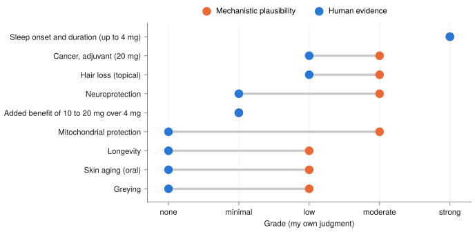
- Quality is low where the claims are largest. The cancer trials are small and mostly high risk of bias; the Parkinson's signal is a subgroup of five trials; the hair trial is a pilot of 40 women.3,11,15
- Most mechanisms are preclinical and at high concentrations. Mouse, rat and cell work dominates the mitochondrial literature, and human tissue pharmacokinetics are essentially unmeasured.
- Several statements here are background knowledge. Half-life, bioavailability, CYP1A2 effects, the phase-response curve and the scavenging-kinetics argument were not retrieved from a source, and the Sources note says so.
- Searches were not exhaustive. Skin and greying searches were noisy, so I may have missed trials.
For sleep, the evidence supports a few milligrams taken early, and a larger immediate-release dose mostly lengthens exposure rather than strengthening it. Beyond sleep, the biology is credible and coherent, mitochondria most of all, but the evidence is lopsided: strong in cells and rodents, thin in people, and tied to concentrations an oral dose may not reach. I would treat everything past sleep as upside you cannot count on, and the strongest remaining human signal, adjunctive cancer care, as a question for an oncologist and not a supplement decision.
Sources
- Cruz-Sanabria F, Bruno S, Crippa A, et al. Optimizing the time and dose of melatonin as a sleep-promoting drug: a systematic review of randomized controlled trials and dose-response meta-analysis. J Pineal Res 76:e12985, 2024. doi:10.1111/jpi.12985
- Bruno S, Cenerini G, Lo Giudice L, et al. Optimizing timing and dose of exogenous melatonin administration in neuropsychiatric pediatric populations: a meta-analysis on sleep outcomes. Sleep Med Rev 84:102158, 2025. doi:10.1016/j.smrv.2025.102158
- Zrineh A, Akwan R, Elsharkawy MM, et al. The effect of melatonin on sleep quality and daytime sleepiness in Parkinson's disease: a systematic review and meta-analysis of randomized placebo-controlled trials. Sleep Med 131:106540, 2025. doi:10.1016/j.sleep.2025.106540
- Nabil Y, Ghazou A, Abodabesh S, et al. Melatonin supplementation in multiple sclerosis: a systematic review of clinical, imaging, biomarker, and safety outcomes. Front Neurol 17:1889519, 2026. doi:10.3389/fneur.2026.1889519
- Dupré C, Legros C, Boutin JA. Functionality of melatonin receptors: recruitment of β-arrestin at MT1. Methods Mol Biol 2550:195–199, 2022. doi:10.1007/978-1-0716-2593-4_24
- Boutin JA, Hamon de Almeida V, Coussay N, et al. Melatonin facts: melatonin lacks immuno-inflammation boosting capacities at the molecular and cellular levels. Biochimie 222:195–202, 2024. doi:10.1016/j.biochi.2024.03.010
- Mathes AM, Heymann P, Ruf C, et al. Endogenous and exogenous melatonin exposure attenuates hepatic MT1 melatonin receptor protein expression in rat. Antioxidants 8(9):408, 2019. doi:10.3390/antiox8090408
- Lin CC, Hou YC, Hsiao PJ, Lu KC. The circadian-melatonin axis in bone remodeling: receptor-dependent signaling, receptor-independent actions, and translational constraints. Biomolecules 16(9):1232, 2026. doi:10.3390/biom16091232
- Wang YM, et al. The efficacy and safety of melatonin in concurrent chemotherapy or radiotherapy for solid tumors: a meta-analysis of randomized controlled trials. Cancer Chemother Pharmacol, 2012. doi:10.1007/s00280-012-1828-8
- Seely D, Wu P, Fritz H, et al. Melatonin as adjuvant cancer care with and without chemotherapy: a systematic review and meta-analysis of randomized trials. Integr Cancer Ther, 2012. doi:10.1177/1534735411425484
- Yu ZY, Peng RY, et al. Melatonin in cancer treatment. Cochrane Database Syst Rev, 2025. doi:10.1002/14651858.CD010145.pub2
- Fan R, et al. Effect of melatonin on quality of life and symptoms in patients with cancer: a systematic review and meta-analysis. BMJ Open, 2022. doi:10.1136/bmjopen-2022-060912
- Veiga ECA, et al. Repercussions of melatonin on the risk of breast cancer: a systematic review and meta-analysis. Rev Assoc Med Bras, 2019. doi:10.1590/1806-9282.65.5.699
- Lissoni P, Tancini G, et al. Treatment of cancer chemotherapy-induced toxicity with the pineal hormone melatonin. Support Care Cancer, 1997. doi:10.1007/BF01262569
- Fischer TW, et al. Melatonin increases anagen hair rate in women with androgenetic alopecia or diffuse alopecia: results of a pilot randomized controlled trial. Br J Dermatol, 2004. doi:10.1111/j.1365-2133.2004.05685.x
- Babadjouni A, Reddy M, Zhang R, Raffi J, et al. Melatonin and the human hair follicle. J Drugs Dermatol 22(3), 2023. doi:10.36849/JDD.6921
- Niu YL, Li YK, et al. Melatonin promotes hair regeneration by modulating the Wnt/β-catenin signalling pathway. Cell Prolif, 2024. doi:10.1111/cpr.13656
- Slominski AT, et al. Melatonin: a cutaneous perspective on its production, metabolism, and functions. J Invest Dermatol, 2018. doi:10.1016/j.jid.2017.10.025
- Bocheva G, Bakalov D, et al. The vital role of melatonin and its metabolites in the neuroprotection and retardation of brain aging. Int J Mol Sci, 2024. doi:10.3390/ijms25105122
- Huo X, Wang C, et al. Human transporters, PEPT1/2, facilitate melatonin transportation into mitochondria of cancer cells: an implication of the therapeutic potential. J Pineal Res, 2017. doi:10.1111/jpi.12390
- Suofu Y, et al. Dual role of mitochondria in producing melatonin and driving GPCR signaling to block cytochrome c release. PNAS, 2017. doi:10.1073/pnas.1705768114
- Zhang Y, Cook A, Kim J, et al. Melatonin inhibits the caspase-1/cytochrome c/caspase-3 cell death pathway, inhibits MT1 receptor loss and delays disease progression in a mouse model of amyotrophic lateral sclerosis. Neurobiol Dis, 2013. doi:10.1016/j.nbd.2013.03.008
- Radogna F, Cristofanon S, Paternoster L, et al. Melatonin antagonizes the intrinsic pathway of apoptosis via mitochondrial targeting of Bcl-2. J Pineal Res, 2008. doi:10.1111/j.1600-079X.2007.00532.x
- Fang Y, et al. Melatonin improves cryopreservation of ram sperm by inhibiting mitochondrial permeability transition pore opening. Reprod Domest Anim, 2020. doi:10.1111/rda.13771
- Jia P, Liu C, et al. Agomelatine protects against myocardial ischemia reperfusion injury by inhibiting mitochondrial permeability transition pore opening. Am J Transl Res, 2018 (no DOI listed).
- Niu FW, Liu MD, et al. Mitochondrial ROS-associated integrated stress response is involved in arsenic-induced blood-testis barrier disruption and protective effect of melatonin. Environ Int, 2025. doi:10.1016/j.envint.2025.109346
- Gao L, Shen J, et al. Melatonin ameliorates decaBDE-induced autism-relevant behaviors through promoting SIRT1/SIRT3/FOXO3a-dependent mitochondrial quality control. Antioxidants 15:405, 2026. doi:10.3390/antiox15030405
- Yürüker V, Nazıroğlu M, et al. Reduction in traumatic brain injury-induced oxidative stress, apoptosis, and calcium entry in rat hippocampus by melatonin: possible involvement of TRPM2 channels. Metab Brain Dis, 2014. doi:10.1007/s11011-014-9623-3
- Peres MFP, Zukerman E, et al. Melatonin, 3 mg, is effective for migraine prevention. Neurology, 2004. doi:10.1212/01.wnl.0000134653.35587.24
What I checked. References 1 to 29 were retrieved from PubMed and I worked from their abstracts and metadata, not the full texts, so effect sizes are as the abstracts report them. First authors, journals and years come from the PubMed records; volume and page numbers are omitted where I did not retrieve them. Reference 25 has no DOI in its record. Reference 14 is a 1997 pilot trial. A 2005 critical appraisal of reference 15 exists, but its abstract was unavailable to me and I have not read its argument. Reference 8 is a bone-remodeling review and reference 6 is an inflammation study; I apply their exposure argument to other outcomes by analogy.
What is from background knowledge, not retrieved. The half-life of about 45 minutes, oral bioavailability of about 15%, CYP1A2 variance and inhibitors, the phase-response curve and morning-overlap argument, the plasma concentration ranges in Figure 3, the kinetic argument that direct scavenging cannot compete with SOD2 and glutathione peroxidase, topical UV-erythema data, the human glucose-tolerance signal, IARC night-shift classification, and rodent dosing ranges. I searched for a paper making the kinetic argument and did not find one. Figures 1 and 2 are illustrative models, and the grades in Figure 5 are my own judgment.
Scope. This review has not been peer reviewed and no original data were collected. It is a research review, not medical advice.